Abstract
Case Report
Clinical, histopathological and surgical evaluations of persistent oropharyngeal membrane case in a calf
Vehbi Gunes*, Gultekin Atalan, Latife Cakir Bayram, Kemal Varol, Hanifi Erol, Ihsan Keles and Ali C Onmaz
Published: 05 August, 2019 | Volume 3 - Issue 1 | Pages: 021-025
A male, 4 days old and 20 kg Simmental calf was evaluated for regurgitation and hyper salivation since birth. The mother became pregnant by artificial insemination and the pregnancy was the second of the mother. A membrane closed the pharynx and a diverticulum on dorsal of this membrane was seen during oropharyngeal examination through inspection. Membrane was also viewed by endoscopy under general anaesthesia. Larynx and oesophagus were imaged by bronchoscopy through the back side of the membrane. After these applications, it was decided that soft palate adhered firmly to the root of tongue causing congenital atresia. Surgical treatment of oropharyngeal membrane was carried out under general anaesthesia. Firstly, tracheotomy was performed for to ease breathing and membrane removed by electrocautery application. Intensive fluid accumulation and oedema formation at the incision area were detected by endoscopic examination following operation and the calf had severe dyspnoea two days after operation and died due to respiratory insufficiency. At necropsy, severe inflammatory reaction, laryngeal oedema and intensive salivation at the surgical side was determined. Direct imaging techniques should be used to determine in the closed oropharyngeal lumen. Moreover, nasopharyngoscopy should be considered to image larynx and oesophageal way. Present case is the first report with concern to pharyngeal membrane formation together with direct imaging and surgical procedures. Therefore, it was considered that this case report could be useful for colleagues and literatures.
Read Full Article HTML DOI: 10.29328/journal.acr.1001016 Cite this Article Read Full Article PDF
Keywords:
Calf; Persistent oropharyngeal membrane; Anomaly
References
- x Hyttel P, Sinowatz F, Vejlsted M, Betteridge K: Essentials of domestic animal embryology. Saunders, Elsevier. 2009, 246-304.
- McGeady TA, Quinn PJ, Fitz Patrick ES, Ryan MT. Veterinary embryology. John Wiley & Sons, Blackwell Publishing. 2013, 205-287.
- Ramachandran S, Panchaksharappa MG, Annigeri RG, Shettar SS. Persistent buccopharyngeal membrane in an adult an incidental finding–A case report. Int j dent clin. 2010; 2: 74-76.
- Verma S, Geller K. Persistent buccopharyngeal membrane: Report of a case and review of the literature. Int J Pediatr Otorhinolaryngol 2009; 73: 877-880.
- Tan S, Tan H, Lim C, Chiang W. A novel stent for the treatment of persistent buccopharyngeal membrane. Int J Pediatr Otorhinolaryngol. 2006; 70: 1645-1649.
- Kara C, Kara I. Persistent buccopharyngeal membrane. Otolaryngol Head Neck Surg. 2007; 136: 1021-1022. PubMed: https://www.ncbi.nlm.nih.gov/pubmed/17548001
- Latshaw WK. Veterinary Developmental Anatomy. BC Decker, Philadelphia, PA. 1987; 87-126.
- Smoak IW, Hudson LC. Persistent oropharyngeal membrane in a Hereford calf. Vet Pathol 1996; 33: 80-82. PubMed: https://www.ncbi.nlm.nih.gov/pubmed/8826010
- Ooi EH, Khouri Z, Hilton M. Persistent buccopharyngeal membrane in an adult. Int J Oral Maxillofac Surg. 2005; 34: 446-448. PubMed: https://www.ncbi.nlm.nih.gov/pubmed/16053859
- Fubini SL, Ducharme N. Farm animal surgery. Elsevier Health Sciences, 2004, 226.
- Aiello SE. Hematologic Reference Ranges. Merck Sharp & Dohme Corp., a subsidiary of Merck & Co., Inc., Kenilworth, NJ., USA. 2017; http://www.merckvetmanual.com/mvm/appendixes/reference_guides/hematologic_reference_ranges.html#v3362733.
- Longacre JJ. Congenital atresia of the oropharynx. Plast Reconstr Surg (1946). 1951; 8: 341-348. PubMed: https://www.ncbi.nlm.nih.gov/pubmed/14891381
- Klinkon M, Ježek J. Values of blood variables in calves. In: A Bird’s-Eye View of Veterinary Medicine. Slovenia, Intech. 2012; 301-320.
- Brun Hansen HC, Kampen AH, Lund A. Hematologic values in calves during the first 6 months of life. Vet Clin Pathol 2006; 35: 182-187. PubMed: https://www.ncbi.nlm.nih.gov/pubmed/16783710
- Marutsova V, Binev R, Marutsov P. Comparative Clinical and Haematological Investigations in Lactating Cows with Subclinical and Clinical Ketosis. Mac Vet Rev. 2015, 38: 2159-2166.
- Panousis N, Siachos N, Kitkas G, Kalaitzakis E, Kritsepi-Konstantinou M, et al. Hematology reference intervals for neonatal Holstein calves. Res Vet Sci. 2018; 118: 1-10. PubMed: https://www.ncbi.nlm.nih.gov/pubmed/29331737
Figures:

Figure 1
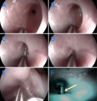
Figure 2

Figure 3
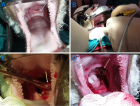
Figure 4
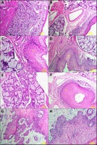
Figure 5
Similar Articles
-
Clinical, histopathological and surgical evaluations of persistent oropharyngeal membrane case in a calfVehbi Gunes*,Gultekin Atalan,Latife Cakir Bayram,Kemal Varol,Hanifi Erol,Ihsan Keles,Ali C Onmaz. Clinical, histopathological and surgical evaluations of persistent oropharyngeal membrane case in a calf. . 2019 doi: 10.29328/journal.acr.1001016; 3: 021-025
-
Complex cyanotic congenital heart disease presenting as congenital heart block in a Nigerian infant: case report and literature reviewUjuanbi Amenawon Susan*,Amain Ebidimie Divine,Gregory Frances. Complex cyanotic congenital heart disease presenting as congenital heart block in a Nigerian infant: case report and literature review. . 2022 doi: 10.29328/journal.acr.1001058; 6: 009-012
-
Lymphoscintigraphic Investigations for Women with Lower Limb Edemas After One PregnancyCallebaut Gregory,Leduc Olivier,Pierre Bourgeois*. Lymphoscintigraphic Investigations for Women with Lower Limb Edemas After One Pregnancy. . 2025 doi: 10.29328/journal.acr.1001129; 9: 066-072
Recently Viewed
-
Sinonasal Myxoma Extending into the Orbit in a 4-Year Old: A Case PresentationJulian A Purrinos*, Ramzi Younis. Sinonasal Myxoma Extending into the Orbit in a 4-Year Old: A Case Presentation. Arch Case Rep. 2024: doi: 10.29328/journal.acr.1001099; 8: 075-077
-
High-Performance Liquid Chromatography (HPLC): A reviewAbdu Hussen Ali*. High-Performance Liquid Chromatography (HPLC): A review. Ann Adv Chem. 2022: doi: 10.29328/journal.aac.1001026; 6: 010-020
-
Drug Rehabilitation Centre-based Survey on Drug Dependence in District Shimla Himachal PradeshKanishka Saini,Palak Sharma,Bhawna Sharma*,Atul Kumar Dubey,Muskan Bhatnoo,Prajkta Thakur,Vanshika Chandel,Ritika Sinha. Drug Rehabilitation Centre-based Survey on Drug Dependence in District Shimla Himachal Pradesh. J Addict Ther Res. 2025: doi: 10.29328/journal.jatr.1001032; 9: 001-006
-
Poly-dopamine-Beta-Cyclodextrin Modified Glassy Carbon Electrode as a Sensor for the Voltammetric Detection of L-Tryptophan at Physiological pHMohammad Hasanzadeh*,Nasrin Shadjou,Sattar Sadeghi,Ahad Mokhtarzadeh,Ayub karimzadeh. Poly-dopamine-Beta-Cyclodextrin Modified Glassy Carbon Electrode as a Sensor for the Voltammetric Detection of L-Tryptophan at Physiological pH . J Forensic Sci Res. 2017: doi: 10.29328/journal.jfsr.1001001; 1: 001-009
-
WMW: A Secure, Web based Middleware for C4I Interoperable ApplicationsNida Zeeshan*. WMW: A Secure, Web based Middleware for C4I Interoperable Applications. J Forensic Sci Res. 2017: doi: 10.29328/journal.jfsr.1001002; 1: 010-017
Most Viewed
-
Evaluation of Biostimulants Based on Recovered Protein Hydrolysates from Animal By-products as Plant Growth EnhancersH Pérez-Aguilar*, M Lacruz-Asaro, F Arán-Ais. Evaluation of Biostimulants Based on Recovered Protein Hydrolysates from Animal By-products as Plant Growth Enhancers. J Plant Sci Phytopathol. 2023 doi: 10.29328/journal.jpsp.1001104; 7: 042-047
-
Sinonasal Myxoma Extending into the Orbit in a 4-Year Old: A Case PresentationJulian A Purrinos*, Ramzi Younis. Sinonasal Myxoma Extending into the Orbit in a 4-Year Old: A Case Presentation. Arch Case Rep. 2024 doi: 10.29328/journal.acr.1001099; 8: 075-077
-
Feasibility study of magnetic sensing for detecting single-neuron action potentialsDenis Tonini,Kai Wu,Renata Saha,Jian-Ping Wang*. Feasibility study of magnetic sensing for detecting single-neuron action potentials. Ann Biomed Sci Eng. 2022 doi: 10.29328/journal.abse.1001018; 6: 019-029
-
Pediatric Dysgerminoma: Unveiling a Rare Ovarian TumorFaten Limaiem*, Khalil Saffar, Ahmed Halouani. Pediatric Dysgerminoma: Unveiling a Rare Ovarian Tumor. Arch Case Rep. 2024 doi: 10.29328/journal.acr.1001087; 8: 010-013
-
Physical activity can change the physiological and psychological circumstances during COVID-19 pandemic: A narrative reviewKhashayar Maroufi*. Physical activity can change the physiological and psychological circumstances during COVID-19 pandemic: A narrative review. J Sports Med Ther. 2021 doi: 10.29328/journal.jsmt.1001051; 6: 001-007

HSPI: We're glad you're here. Please click "create a new Query" if you are a new visitor to our website and need further information from us.
If you are already a member of our network and need to keep track of any developments regarding a question you have already submitted, click "take me to my Query."


