Abstract
Case Report
Scraping cytology and scanning electron microscopy in diagnosis and therapy of corneal ulcer by mycobacterium infection
Russo Giacomo*, Del Prete Salvatore, Del Prete Antonio, Meloni Marisa, Capaldi Roberto and Grumetto Lucia
Published: 06 December, 2019 | Volume 3 - Issue 1 | Pages: 050-053
Purpose: This work is aimed at demonstrating that scraping cytology and scanning electron microscopy can successfully assist in the diagnosis of nontuberculous mycobacteria infection. For this purpose, we report the use of both these techniques in the diagnosis of cornel ulcer in a previously healthy young man.
Methods: Cytological samples were achieved by scraping technique on the mucosa, both sub palpebral and temporal area of the eye tarsal conjunctiva. The obtained sample was affixed to a sanded rectangular slide, stained with the Pappenheim method, washed in bidistilled water, treated in Giemsa solution, washed again and subsequently dried on a hot plate and observed with a microscope at various magnifications.
Results: After a therapy based on a 500 mg clarithromycin tablet administered every 12 hours for 30 days as systemic therapy, a complete recovery of the patient from left eye inflammation was observed and SEM cytology showed that NTM colonies had disappeared.
Conclusion: Conjunctival cytology scraping and SEM technologies can be therefore exploited as new tools in diagnosis and fast identification of these newly discovered mycobacteria. In fact, they have a new way for studying ocular pathology, because of the simple execution and remarkable accuracy in the diagnosis. In fact, this technique allows to gather valuable information about all pathogens expression and the cellular action involved in pathology. As a further plus, this technique provides clinicians with the opportunity to repeat the SEM cytology for monitoring patients during therapy, hence leading to evaluate the efficacy of the pharmaceutical regimen in real time.
Read Full Article HTML DOI: 10.29328/journal.acr.1001024 Cite this Article Read Full Article PDF
References
- Henkle E, Winthrop K. Nontuberculous Mycobacteria Infections in Immunosuppressed. Hosts. Clin Chest Med. 2015; 36: 91–99. PubMed: https://www.ncbi.nlm.nih.gov/pubmed/25676522
- org.https://www.thoracic.org/statements/resources/mtpi/nontuberculous-mycobacterial-diseases.pdf.
- Henry CR, Flynn HW, Mille D, Forster RK, Alfonso EC. Infectious keratitis progressing to endophthalmitis: a 15-year study of microbiology, associated factors, and clinical outcomes. Ophthalmology. 2012; 119: 2443-2449. PubMed: https://www.ncbi.nlm.nih.gov/pubmed/22858123
- Hernandez-Toloza JE, Rincon-Serrano Mde P, Celis-Bustos YA, Aguillon CI. Identification of mycobacteria to the species level by molecular methods in the Public Health Laboratory of Bogota, Colombia. Enferm Infecc Microbiol Clin. 2016; 34: 17-22.
- Yam WC, Siu KH. Rapid identification of mycobacteria and rapid detection of drug resistance in Mycobacterium tuberculosis in cultured isolates and in respiratory specimens. Methods Mol Biol. 2013; 943: 171-99. PubMed: https://www.ncbi.nlm.nih.gov/pubmed/23104290
- Morgan MA, Doerr KA, Hempel HO, Goodman NL, Roberts GD. Evaluation of the p-nitro-alpha-acetylamino-beta-hydroxypropiophenone differential test for identification of Mycobacterium tuberculosis complex. J Clin Microbiol 1985; 21: 634-635. PubMed: https://www.ncbi.nlm.nih.gov/pmc/articles/PMC271735/
- Cennamo GL, Del Prete A, Forte R, Cafiero G, Del Prete S, et al. Impression cytology with scanning electron microscopy: a new method in the study of conjunctival microvilli. Eye. 2008; 22: 138-143. PubMed: https://www.ncbi.nlm.nih.gov/pubmed/17603470
- Golding CG, Lamboo LL, Beniac DR, Booth TF. The scanning electron microscope in microbiology and diagnosis of infectious disease. Sci Rep. 2016; 6: 26516. PubMed: https://www.ncbi.nlm.nih.gov/pubmed/27212232
- Carter HW. Clinical applications of scanning electron microscopy (SEM) in North America with emphasis on SEM's role in comparative microscopy. Scan Electron Microsc. 1980; 115-120. PubMed: https://www.ncbi.nlm.nih.gov/pubmed/7414259
- Set R, Shastri J. Laboratory aspects of clinically significant rapidly growing mycobacteria. Indian J Med Microbiol. 2011; 29: 343-352. PubMed: https://www.ncbi.nlm.nih.gov/pubmed/22120792
- Bittner MJ, Preheim LC. Other Slow-Growing Nontuberculous Mycobacteria. Microbiol Spectr. 2016; 4. PubMed: https://www.ncbi.nlm.nih.gov/pubmed/27837745
Figures:
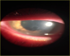
Figure 1
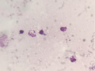
Figure 2
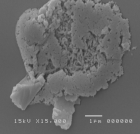
Figure 3
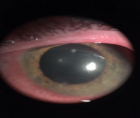
Figure 4

Figure 5
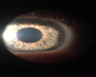
Figure 6
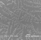
Figure 7
Similar Articles
-
Trichomonas Vaginalis-A Clinical ImageAstrit M Gashi*. Trichomonas Vaginalis-A Clinical Image. . 2017 doi: 10.29328/journal.hjcr.1001001; 1: 001-002
-
Gastric Mucosal CalcinosisVedat Goral*,Irem Ozover,Ilknur Turkmen. Gastric Mucosal Calcinosis. . 2017 doi: 10.29328/journal.hjcr.1001002; 1: 003-005
-
Giant Lipoma Anterior Neck: A case reportManu CB*,Gowri Sankar M,Arun Alexander. Giant Lipoma Anterior Neck: A case report . . 2017 doi: 10.29328/journal.hjcr.1001003; 1: 006-008
-
Catamenial pneumothorax: Presentation of an uncommon PathologyRui Haddad*,Caterin Arévalo,David Nigri. Catamenial pneumothorax: Presentation of an uncommon Pathology . . 2017 doi: 10.29328/journal.hjcr.1001004; 1: 009-013
-
A rare case: Congenital Megalourethra in prune belly syndromeNuman Baydilli*,Ismail Selvi,Emre Can Akınsal. A rare case: Congenital Megalourethra in prune belly syndrome. . 2018 doi: 10.29328/journal.acr.1001005; 2: 001-003
-
Trauma to the neck: Manifestation of injuries outside the original zone of injury-A case reportStephen O Heard*,Alexander Christakis,Brian Tashjian,Anne M Gilroy3. Trauma to the neck: Manifestation of injuries outside the original zone of injury-A case report. . 2018 doi: 10.29328/journal.acr.1001006; 2: 004-009
-
Meige Trofoedema: A form of primary lymphedemaCarlos Al Sanchez Salguero*. Meige Trofoedema: A form of primary lymphedema. . 2018 doi: 10.29328/journal.acr.1001007; 2: 010-014
-
A rare case of Diabetic Foot in male of middle age has been shown Diabetic footSujit K Bhattacharya*. A rare case of Diabetic Foot in male of middle age has been shown Diabetic foot. . 2018 doi: 10.29328/journal.acr.1001008; 2: 015-015
-
Brooke-Spiegler Syndrome: A rare cause of skin appendageal tumorN Suganthan*,S Pirasath,DD Dikowita. Brooke-Spiegler Syndrome: A rare cause of skin appendageal tumor. . 2018 doi: 10.29328/journal.acr.1001009; 2: 016-018
-
McArdle’s Disease (Glycogen Storage Disease type V): A Clinical CaseCameselle-Teijeiro JF*,Calheiros-Cruz T,Caamaño-Vara MP,Villar-Fernández B,Ruibal-Azevedo J,Cameselle-Cortizo L,Cameselle-Arias M,Charro Gamallo ME,Turienzo-Pacho F,Yera Acosta A. McArdle’s Disease (Glycogen Storage Disease type V): A Clinical Case. . 2018 doi: 10.29328/journal.acr.1001010; 2: 019-023
Recently Viewed
-
Fostering Pathways and Creativity Responsible for Advancing Health Research Skills and Knowledge for Healthcare Professionals to Heighten Evidence-Based Healthcare Practices in Resource-Constrained Healthcare SettingsJanvier Nzayikorera*. Fostering Pathways and Creativity Responsible for Advancing Health Research Skills and Knowledge for Healthcare Professionals to Heighten Evidence-Based Healthcare Practices in Resource-Constrained Healthcare Settings. J Community Med Health Solut. 2025: doi: 10.29328/journal.jcmhs.1001052; 6: 005-019
-
Chaos to Cosmos: Quantum Whispers and the Cosmic GenesisOwais Farooq*,Romana Zahoor*. Chaos to Cosmos: Quantum Whispers and the Cosmic Genesis. Int J Phys Res Appl. 2025: doi: 10.29328/journal.ijpra.1001107; 8: 017-023
-
Buffer Solutions of known Ionic StrengthVíctor Cerdà*, Piyawan Phansi. Buffer Solutions of known Ionic Strength. Ann Adv Chem. 2023: doi: 10.29328/journal.aac.1001043; 7: 051-056
-
Sinonasal Myxoma Extending into the Orbit in a 4-Year Old: A Case PresentationJulian A Purrinos*, Ramzi Younis. Sinonasal Myxoma Extending into the Orbit in a 4-Year Old: A Case Presentation. Arch Case Rep. 2024: doi: 10.29328/journal.acr.1001099; 8: 075-077
-
Comparative Study of Cerebral Volumetric Variations in Patients with Schizophrenia with their Unaffected First-degree Relatives, using Magnetic Resonance Imaging Technique, a Case-control StudyMahdiye Fanayi,Mohammad Ali Oghabian*,Hamid Reza Naghavi,Hassan Farrahi. Comparative Study of Cerebral Volumetric Variations in Patients with Schizophrenia with their Unaffected First-degree Relatives, using Magnetic Resonance Imaging Technique, a Case-control Study. J Neurosci Neurol Disord. 2024: doi: 10.29328/journal.jnnd.1001088; 8: 001-007
Most Viewed
-
Evaluation of Biostimulants Based on Recovered Protein Hydrolysates from Animal By-products as Plant Growth EnhancersH Pérez-Aguilar*, M Lacruz-Asaro, F Arán-Ais. Evaluation of Biostimulants Based on Recovered Protein Hydrolysates from Animal By-products as Plant Growth Enhancers. J Plant Sci Phytopathol. 2023 doi: 10.29328/journal.jpsp.1001104; 7: 042-047
-
Sinonasal Myxoma Extending into the Orbit in a 4-Year Old: A Case PresentationJulian A Purrinos*, Ramzi Younis. Sinonasal Myxoma Extending into the Orbit in a 4-Year Old: A Case Presentation. Arch Case Rep. 2024 doi: 10.29328/journal.acr.1001099; 8: 075-077
-
Feasibility study of magnetic sensing for detecting single-neuron action potentialsDenis Tonini,Kai Wu,Renata Saha,Jian-Ping Wang*. Feasibility study of magnetic sensing for detecting single-neuron action potentials. Ann Biomed Sci Eng. 2022 doi: 10.29328/journal.abse.1001018; 6: 019-029
-
Pediatric Dysgerminoma: Unveiling a Rare Ovarian TumorFaten Limaiem*, Khalil Saffar, Ahmed Halouani. Pediatric Dysgerminoma: Unveiling a Rare Ovarian Tumor. Arch Case Rep. 2024 doi: 10.29328/journal.acr.1001087; 8: 010-013
-
Physical activity can change the physiological and psychological circumstances during COVID-19 pandemic: A narrative reviewKhashayar Maroufi*. Physical activity can change the physiological and psychological circumstances during COVID-19 pandemic: A narrative review. J Sports Med Ther. 2021 doi: 10.29328/journal.jsmt.1001051; 6: 001-007

HSPI: We're glad you're here. Please click "create a new Query" if you are a new visitor to our website and need further information from us.
If you are already a member of our network and need to keep track of any developments regarding a question you have already submitted, click "take me to my Query."


