Case Report
Giant Lipoma Anterior Neck: A case report

Gowri Sankar M1, Manu CB2* and Arun Alexander3
1Senior Resident, Department of ENT, JIPMER, India
2Junior Resident, Department of ENT, JIPMER, India
3Additional Professor and Head, Department of ENT, JIPMER, India
*Address for Correspondence: Manu CB, Junior Resident, Department of ENT, JIPMER, India, Email: cbalakrishnanmanu@gmail.com
Dates: Submitted: 06 November 2017; Approved: 12 December 2017; Published: 14 December 2017
How to cite this article: Gowri Sankar M, Manu CB, Alexander A. Giant Lipoma Anterior Neck: A case report. Arch Case Rep. 2017; 1: 006-008. DOI: 10.29328/journal.acr.1001003
Copyright License: © 2017 Gowri Sankar M, et al. This is an open access article distributed under the Creative Commons Attribution License, which permits unrestricted use, distribution, and reproduction in any medium, provided the original work is properly cited.
Keywords: Lipoma; Giant lipoma; Anterior neck swelling MeSH terms: Lipoma; Neck
Abstract
Introduction
Lipoma is a benign mesenchymal tumor composed of adult fat cells. Often called as “universal tumor” or an “ubiquitous tumor” as they can occur anywhere in the body where there is accumulation of fat cells [1]. The incidence in head and neck region being thirteen percent [2]. Lipomas in the neck usually involve the posterior triangle. Anterior neck lipomas are a rare entity while giant anterior neck lipomas (>10 cm) are even rarer, we here present one such case [3].
Case Report
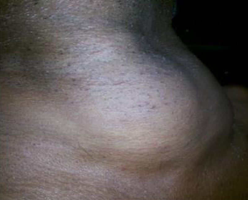
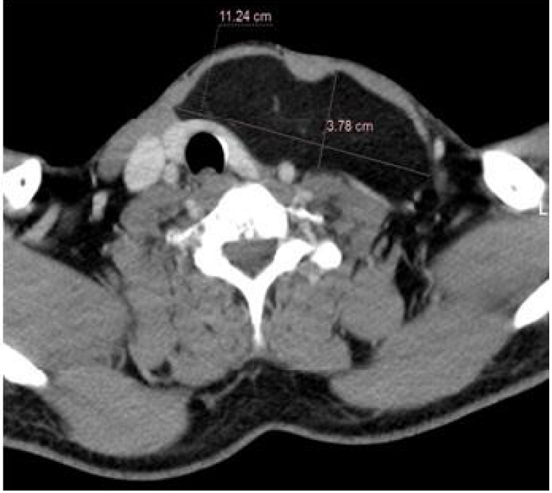
A 50 yr man presented with complains of swelling in the anterior neck for 15 yrs. The swelling appeared to growing rapidly for the last 6 months. He had no other complaints. On examination a swelling of 14×10 cm involving both sides of the anterior neck extending superiorly from the upper ZDFSVVVXV thyroid cartilage to clavicle inferiorly, soft in consistency, freely mobile in all directions of with no movement on deglution. FNAC was suggestive of lipoma (Figure 1). CT showed the lesion measuring 11.4×3.8 cm in the subplatysmal plane with mild displacement of trachea with no compression (Figure 2).
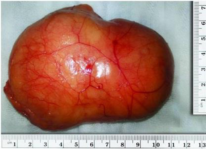
The patient was planned for excision under general anesthesia via routine thyroidectomy incision. The tumor was located beneath the platysma, once a plane was identified between the platysma, tumor and underlying strap muscles, the entire tumor was delivered in toto by finger dissection and “squeeze technique”. Wound was closed in layers and skin with subcuticular sutures. Histopathology of the specimen removed was lipoma (Figure 3). The patient had a cosmetically acceptable scar on the neck and was followed up for 1 year with no recurrence.
Discussion
Lipomas are benign adipose tumors of mesenchymal origin secondary to hamartomatous proliferation of mature fat cells [4]. Lipomas are classified as subcutaneous type, subfascial type or intermuscular type [5]. Cheek is the most favored site in head and neck region followed by the tongue, floor of the mouth, buccal sulcus, vestibule, lip, palate and gingiva rarely occurring subcutaneously in the anterior neck region [3] as was in our case [6]. The exact cause for lipoma is unclear though there is an association with genetic mutation in chromosome 12 in cases of solitary lipomas [7].
Clinical features vary greatly depending upon the lesion’s size, location and rate of growth. Like most benign tumors they present as a painless, mobile, palpable masses which are often overlooked by patients till they become an appreciable mass as was in our case [8]. Giant lipomas measure at least >10cm in one dimension, or weighing at least 1000gm [9]. A rapid increase in size should always raise the suspicion of malignancy. Ultrasonography remains as the initial imaging modality in diagnosis of head and neck lipomas while Fine needle aspiration cytology (FNAC) or computed tomography (CT) is indicated for confirmation of diagnosis [10].
Consecutive follow up might be a valid option for asymptomatic patients with anterior neck lipomas. Surgical intervention for giant lipomas of the anterior neck is challenging, requiring thorough anatomy of the region and meticulous skills given their relationship with vital structures in the anterior neck and should be reserved for patients with cosmesis and pressure effects. Complete excision with capsule should be performed to prevent recurrence.
Conclusion
Lipomas in the anterior neck are rare, but can present as giant lipomas as in our case. There is a paucity of published analogous cases in our literature, hence we conclusively propose to include giant lipomas in the differential diagnosis of swellings of the anterior neck. Adequate preoperative imaging and a good operative technique, surgical excision provides good cosmesis and no functional impairment.
References
- Bailey love_s_short_practice_of_surgery_25th_edition.pdf.
- Leon Barnes. Surgical Pathology of the Head and Neck, Third Edition. Ref.: https://goo.gl/tLXH3P
- Medina CR, Schneider S, Mitra A, Spears J, Mitra A. Giant submental lipoma: Case report and review of the literature. Can J Plast Surg. 2007; 15: 219-222. Ref.: https://goo.gl/dfAm59
- Glick M. Burket’s Oral Medicine. 2015: 732.
- Marx RE, Stern Diane. Oral and Maxillofacial Pathology: A Rationale for Diagnosis and Treatment, Second Edition. Ref.: https://goo.gl/4EQB2W
- Kalia V, Kaushal N, Pahwa D. Giant Subcutaneous Solitary Lipoma Arising In the Neck-Case Report and Review of Literature. 2011; 2: 1-10. Ref.: https://goo.gl/xbDdpC
- Italiano A, Ebran N, Attias R, Chevallier A, Monticelli I, et al. NFIB rearrangement in superficial, retroperitoneal, and colonic lipomas with aberrations involving chromosome band 9p22. Genes Chromosom Cancer. 2008; 47:971-977. Ref.: https://goo.gl/3KusNU
- Del Agua C, Felipo F. Adenolipoma of the skin. Dermatol Online J. 2004; 10: 9. Ref.: https://goo.gl/GAWBjp
- Sanchez MR, Golomb FM, Moy JA, Potozkin JR. Giant lipoma: case report and review of the literature. J Am Acad Dermatol. 1993; 28: 266-268. Ref.: https://goo.gl/NYhZJY
- Sharma BK, Khanna SK, Bharati M, Gupta A. Anterior neck lipoma with anterior mediastinal extention-a rare case report. Kathmandu Univ Med J. 2014; 11: 88-90. Ref.: https://goo.gl/Htzw7g


