Figure 1
Clinical, histopathological and surgical evaluations of persistent oropharyngeal membrane case in a calf
Vehbi Gunes*, Gultekin Atalan, Latife Cakir Bayram, Kemal Varol, Hanifi Erol, Ihsan Keles and Ali C Onmaz
Published: 05 August, 2019 | Volume 3 - Issue 1 | Pages: 021-025
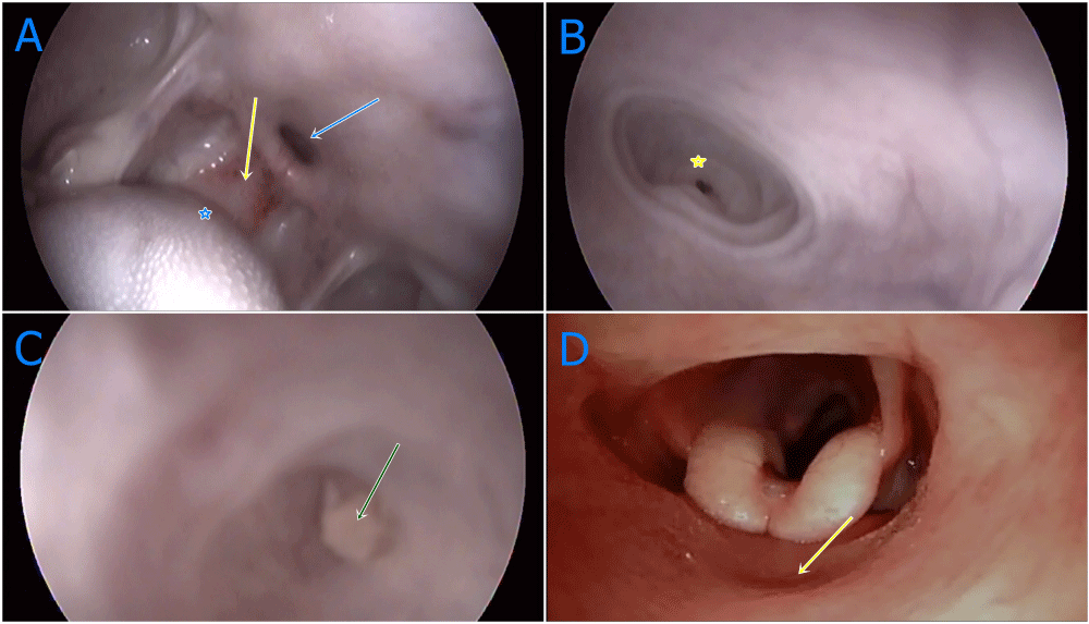
Figure 1:
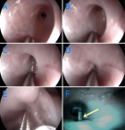
1 A-D: Oropharengeal and nasopharengeal endoscopic images of oropharangeal membrane, A: General appearences of oropharenx with congenital anomaly. Yellow arrow; congenital oropharengeal membran, blue arrow; diverticulum dorsal to oropharengeal membran, blue star; caudal of tonque, B: Near plan image of diverticulum inflated with air. Yellow star; blind sac at the end of diverticulum, C: Coagulated milk accumulation at the end of blind sac (green arrow), D: Broncoscop application via nasal approach, larynx imaging at back of oropharengeal membran (yellow arrow), intermediate wall of oropharengeal membran.
Read Full Article HTML DOI: 10.29328/journal.acr.1001016 Cite this Article Read Full Article PDF
More Images
Similar Articles
-
Clinical, histopathological and surgical evaluations of persistent oropharyngeal membrane case in a calfVehbi Gunes*,Gultekin Atalan,Latife Cakir Bayram,Kemal Varol,Hanifi Erol,Ihsan Keles,Ali C Onmaz. Clinical, histopathological and surgical evaluations of persistent oropharyngeal membrane case in a calf. . 2019 doi: 10.29328/journal.acr.1001016; 3: 021-025
-
Complex cyanotic congenital heart disease presenting as congenital heart block in a Nigerian infant: case report and literature reviewUjuanbi Amenawon Susan*,Amain Ebidimie Divine,Gregory Frances. Complex cyanotic congenital heart disease presenting as congenital heart block in a Nigerian infant: case report and literature review. . 2022 doi: 10.29328/journal.acr.1001058; 6: 009-012
-
Lymphoscintigraphic Investigations for Women with Lower Limb Edemas After One PregnancyCallebaut Gregory,Leduc Olivier,Pierre Bourgeois*. Lymphoscintigraphic Investigations for Women with Lower Limb Edemas After One Pregnancy. . 2025 doi: 10.29328/journal.acr.1001129; 9: 066-072
Recently Viewed
-
Sensitivity and Intertextile variance of amylase paper for saliva detectionAlexander Lotozynski*. Sensitivity and Intertextile variance of amylase paper for saliva detection. J Forensic Sci Res. 2020: doi: 10.29328/journal.jfsr.1001017; 4: 001-003
-
Extraction of DNA from face mask recovered from a kidnapping sceneBassey Nsor*,Inuwa HM. Extraction of DNA from face mask recovered from a kidnapping scene. J Forensic Sci Res. 2022: doi: 10.29328/journal.jfsr.1001029; 6: 001-005
-
The Ketogenic Diet: The Ke(y) - to Success? A Review of Weight Loss, Lipids, and Cardiovascular RiskAngela H Boal*, Christina Kanonidou. The Ketogenic Diet: The Ke(y) - to Success? A Review of Weight Loss, Lipids, and Cardiovascular Risk. J Cardiol Cardiovasc Med. 2024: doi: 10.29328/journal.jccm.1001178; 9: 052-057
-
Could apple cider vinegar be used for health improvement and weight loss?Alexander V Sirotkin*. Could apple cider vinegar be used for health improvement and weight loss?. New Insights Obes Gene Beyond. 2021: doi: 10.29328/journal.niogb.1001016; 5: 014-016
-
Maximizing the Potential of Ketogenic Dieting as a Potent, Safe, Easy-to-Apply and Cost-Effective Anti-Cancer TherapySimeon Ikechukwu Egba*,Daniel Chigbo. Maximizing the Potential of Ketogenic Dieting as a Potent, Safe, Easy-to-Apply and Cost-Effective Anti-Cancer Therapy. Arch Cancer Sci Ther. 2025: doi: 10.29328/journal.acst.1001047; 9: 001-005
Most Viewed
-
Evaluation of Biostimulants Based on Recovered Protein Hydrolysates from Animal By-products as Plant Growth EnhancersH Pérez-Aguilar*, M Lacruz-Asaro, F Arán-Ais. Evaluation of Biostimulants Based on Recovered Protein Hydrolysates from Animal By-products as Plant Growth Enhancers. J Plant Sci Phytopathol. 2023 doi: 10.29328/journal.jpsp.1001104; 7: 042-047
-
Sinonasal Myxoma Extending into the Orbit in a 4-Year Old: A Case PresentationJulian A Purrinos*, Ramzi Younis. Sinonasal Myxoma Extending into the Orbit in a 4-Year Old: A Case Presentation. Arch Case Rep. 2024 doi: 10.29328/journal.acr.1001099; 8: 075-077
-
Feasibility study of magnetic sensing for detecting single-neuron action potentialsDenis Tonini,Kai Wu,Renata Saha,Jian-Ping Wang*. Feasibility study of magnetic sensing for detecting single-neuron action potentials. Ann Biomed Sci Eng. 2022 doi: 10.29328/journal.abse.1001018; 6: 019-029
-
Pediatric Dysgerminoma: Unveiling a Rare Ovarian TumorFaten Limaiem*, Khalil Saffar, Ahmed Halouani. Pediatric Dysgerminoma: Unveiling a Rare Ovarian Tumor. Arch Case Rep. 2024 doi: 10.29328/journal.acr.1001087; 8: 010-013
-
Physical activity can change the physiological and psychological circumstances during COVID-19 pandemic: A narrative reviewKhashayar Maroufi*. Physical activity can change the physiological and psychological circumstances during COVID-19 pandemic: A narrative review. J Sports Med Ther. 2021 doi: 10.29328/journal.jsmt.1001051; 6: 001-007

HSPI: We're glad you're here. Please click "create a new Query" if you are a new visitor to our website and need further information from us.
If you are already a member of our network and need to keep track of any developments regarding a question you have already submitted, click "take me to my Query."


