Figure 1
First cure case of 2019 novel coronavirus in Ningxia, China
Li Zhu*, Yifan Wang, Pan Zhou, Xingcang Tian, Shuping Meng, Xiao Sun and Ting Li
Published: 17 April, 2020 | Volume 4 - Issue 1 | Pages: 026-027
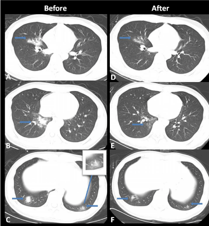
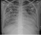
Figure 1:
Thoracic non-contrast CT images in a 28-year-old male before and after treatment. (A-C), Image shows multiple patchy ground-glass opacities and consolidation in the right middle lobe and basal segments of both lower lobes. (D-F), Follow-up image on January 31 shows obviously reduced ground-glass opacities in the right middle lobe and lower lobes of both lungs. The pulmonary vascular thickening is typical CT sign of the 2019 novel coronavirus pneumonia.
Read Full Article HTML DOI: 10.29328/journal.acr.1001035 Cite this Article Read Full Article PDF
More Images
Similar Articles
-
Trichomonas Vaginalis-A Clinical ImageAstrit M Gashi*. Trichomonas Vaginalis-A Clinical Image. . 2017 doi: 10.29328/journal.hjcr.1001001; 1: 001-002
-
Gastric Mucosal CalcinosisVedat Goral*,Irem Ozover,Ilknur Turkmen. Gastric Mucosal Calcinosis. . 2017 doi: 10.29328/journal.hjcr.1001002; 1: 003-005
-
Giant Lipoma Anterior Neck: A case reportManu CB*,Gowri Sankar M,Arun Alexander. Giant Lipoma Anterior Neck: A case report . . 2017 doi: 10.29328/journal.hjcr.1001003; 1: 006-008
-
Catamenial pneumothorax: Presentation of an uncommon PathologyRui Haddad*,Caterin Arévalo,David Nigri. Catamenial pneumothorax: Presentation of an uncommon Pathology . . 2017 doi: 10.29328/journal.hjcr.1001004; 1: 009-013
-
A rare case: Congenital Megalourethra in prune belly syndromeNuman Baydilli*,Ismail Selvi,Emre Can Akınsal. A rare case: Congenital Megalourethra in prune belly syndrome. . 2018 doi: 10.29328/journal.acr.1001005; 2: 001-003
-
Trauma to the neck: Manifestation of injuries outside the original zone of injury-A case reportStephen O Heard*,Alexander Christakis,Brian Tashjian,Anne M Gilroy3. Trauma to the neck: Manifestation of injuries outside the original zone of injury-A case report. . 2018 doi: 10.29328/journal.acr.1001006; 2: 004-009
-
Meige Trofoedema: A form of primary lymphedemaCarlos Al Sanchez Salguero*. Meige Trofoedema: A form of primary lymphedema. . 2018 doi: 10.29328/journal.acr.1001007; 2: 010-014
-
A rare case of Diabetic Foot in male of middle age has been shown Diabetic footSujit K Bhattacharya*. A rare case of Diabetic Foot in male of middle age has been shown Diabetic foot. . 2018 doi: 10.29328/journal.acr.1001008; 2: 015-015
-
Brooke-Spiegler Syndrome: A rare cause of skin appendageal tumorN Suganthan*,S Pirasath,DD Dikowita. Brooke-Spiegler Syndrome: A rare cause of skin appendageal tumor. . 2018 doi: 10.29328/journal.acr.1001009; 2: 016-018
-
McArdle’s Disease (Glycogen Storage Disease type V): A Clinical CaseCameselle-Teijeiro JF*,Calheiros-Cruz T,Caamaño-Vara MP,Villar-Fernández B,Ruibal-Azevedo J,Cameselle-Cortizo L,Cameselle-Arias M,Charro Gamallo ME,Turienzo-Pacho F,Yera Acosta A. McArdle’s Disease (Glycogen Storage Disease type V): A Clinical Case. . 2018 doi: 10.29328/journal.acr.1001010; 2: 019-023
Recently Viewed
-
Kidney Biopsy in Autosomal Dominant Polycystic Kidney DiseaseLaalasa Varanasi*, Gabriel Loeb, Vighnesh Walavalkar, Nebil Mohammed, John Paul Lindsey II, Stephen Gluck, Thomas Lee Chi, Meyeon Park. Kidney Biopsy in Autosomal Dominant Polycystic Kidney Disease. J Clini Nephrol. 2023: doi: 10.29328/journal.jcn.1001118; 7: 101-105
-
What is the Cost of Measuring a Blood Pressure?Steven A Yarows*. What is the Cost of Measuring a Blood Pressure?. Ann Clin Hypertens. 2018: doi: 10.29328/journal.ach.1001012; 2: 059-066
-
Coexistence of common gallstones and sinusoidal obstruction syndrome: Case report and review of the literatureFurkan Karahan*,Nihan Acar,Arzu Avcı,Osman Nuri Dilek. Coexistence of common gallstones and sinusoidal obstruction syndrome: Case report and review of the literature. Arch Surg Clin Res. 2021: doi: 10.29328/journal.ascr.1001060; 5: 020-022
-
The Immunitary role in chronic prostatitis and growth factors as promoter of BPHMauro luisetto*,Behzad Nili-Ahmadabadi,Ghulam Rasool Mashori,Ram Kumar Sahu,Farhan Ahmad Khan,Cabianca luca,Heba Nasser. The Immunitary role in chronic prostatitis and growth factors as promoter of BPH. Insights Clin Cell Immunol. 2018: doi: 10.29328/journal.icci.1001003; 2: 001-013
-
Huge median prostatic lobe: a interesting case of BPHBabty Mouftah*,Slaoui Amine,Fouimtizi Jaafar,Mamad Ayoub,Karmouni Tarik,El Khadder Khalid,Koutani Abdellatif,Ibn Attya Ahmed. Huge median prostatic lobe: a interesting case of BPH. J Clin Med Exp Images. 2022: doi: 10.29328/journal.jcmei.1001026; 6: 006-006
Most Viewed
-
Evaluation of Biostimulants Based on Recovered Protein Hydrolysates from Animal By-products as Plant Growth EnhancersH Pérez-Aguilar*, M Lacruz-Asaro, F Arán-Ais. Evaluation of Biostimulants Based on Recovered Protein Hydrolysates from Animal By-products as Plant Growth Enhancers. J Plant Sci Phytopathol. 2023 doi: 10.29328/journal.jpsp.1001104; 7: 042-047
-
Sinonasal Myxoma Extending into the Orbit in a 4-Year Old: A Case PresentationJulian A Purrinos*, Ramzi Younis. Sinonasal Myxoma Extending into the Orbit in a 4-Year Old: A Case Presentation. Arch Case Rep. 2024 doi: 10.29328/journal.acr.1001099; 8: 075-077
-
Feasibility study of magnetic sensing for detecting single-neuron action potentialsDenis Tonini,Kai Wu,Renata Saha,Jian-Ping Wang*. Feasibility study of magnetic sensing for detecting single-neuron action potentials. Ann Biomed Sci Eng. 2022 doi: 10.29328/journal.abse.1001018; 6: 019-029
-
Pediatric Dysgerminoma: Unveiling a Rare Ovarian TumorFaten Limaiem*, Khalil Saffar, Ahmed Halouani. Pediatric Dysgerminoma: Unveiling a Rare Ovarian Tumor. Arch Case Rep. 2024 doi: 10.29328/journal.acr.1001087; 8: 010-013
-
Physical activity can change the physiological and psychological circumstances during COVID-19 pandemic: A narrative reviewKhashayar Maroufi*. Physical activity can change the physiological and psychological circumstances during COVID-19 pandemic: A narrative review. J Sports Med Ther. 2021 doi: 10.29328/journal.jsmt.1001051; 6: 001-007

HSPI: We're glad you're here. Please click "create a new Query" if you are a new visitor to our website and need further information from us.
If you are already a member of our network and need to keep track of any developments regarding a question you have already submitted, click "take me to my Query."


