Figure 2
Chronic subdural haematoma associated with arachnoid cyst of the middle fossa in a soccer player: Case report and review of the literature
Elena Beretta*, Michele Incerti, Giuseppe Raudino, Gaspare F Montemagno and Franco Servadei
Published: 16 May, 2020 | Volume 4 - Issue 1 | Pages: 032-037
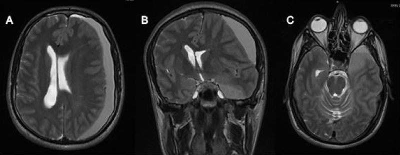
Figure 2:
A) T2 MRI axial view showing a hyperintense left frontotemporal sub-acute hematoma, 20 mm thick with midline shift. B) T2 MRI coronal view showing the subdural hematoma with the same signal og the polar arachnoid cyst. C) T2 MRI axial view showing the communication of the subdural hematoma with the temporal arachnoid cyst.
Read Full Article HTML DOI: 10.29328/journal.acr.1001037 Cite this Article Read Full Article PDF
More Images
Similar Articles
-
Chronic subdural haematoma associated with arachnoid cyst of the middle fossa in a soccer player: Case report and review of the literatureElena Beretta*,Michele Incerti,Giuseppe Raudino,Gaspare F Montemagno,Franco Servadei. Chronic subdural haematoma associated with arachnoid cyst of the middle fossa in a soccer player: Case report and review of the literature. . 2020 doi: 10.29328/journal.acr.1001037; 4: 032-037
Recently Viewed
-
Clinical Case of Successful Therapy for the Patient with Autism by use of Fetal Stem CellsAA Sinelnyk*, SG Shmyh, IG Matiyashchuk, MO Klunnyk, MP Demchuk, OV Ivankova, OO Honza, IA Susak, MV Skalozub, DV Vatlitsov, KI Sorochynska. Clinical Case of Successful Therapy for the Patient with Autism by use of Fetal Stem Cells. J Stem Cell Ther Transplant. 2024: doi: 10.29328/journal.jsctt.1001043; 8: 048-053
-
Surgical Management of Extrahepatic Biliary Neuroendocrine Tumors: A Case ReportNegar Ostadsharif,Alireza Firouzfar,Parto Nasri,Behnam Sanei*. Surgical Management of Extrahepatic Biliary Neuroendocrine Tumors: A Case Report. Arch Case Rep. 2025: doi: 10.29328/journal.acr.1001120; 9: 004-007
-
Hypochlorous acid has emerged as a potential alternative to conventional antibiotics due to its broad-spectrum antimicrobial activityMaher M Akl*. Hypochlorous acid has emerged as a potential alternative to conventional antibiotics due to its broad-spectrum antimicrobial activity. Int J Clin Microbiol Biochem Technol. 2023: doi: 10.29328/journal.ijcmbt.1001026; 6: 001-004
-
KYAMOS Software - Mini Review on the Computer-Aided Engineering IndustryAntonis Papadakis*,Sofia Nikolaidou. KYAMOS Software - Mini Review on the Computer-Aided Engineering Industry. Int J Phys Res Appl. 2025: doi: 10.29328/journal.ijpra.1001105; 8: 010-012
-
Ischemic Stroke and Myocarditis Revealing Behçet’s Disease in a Young Adult: Diagnostic Challenges and Therapeutic PerspectivesObeidat Saleh Muhammed*,Abdallani B,Amine Z,Boucetta A,Bouziane M,Haboub M,Habbal R. Ischemic Stroke and Myocarditis Revealing Behçet’s Disease in a Young Adult: Diagnostic Challenges and Therapeutic Perspectives. J Cardiol Cardiovasc Med. 2025: doi: 10.29328/journal.jccm.1001205; 10: 016-021
Most Viewed
-
Evaluation of Biostimulants Based on Recovered Protein Hydrolysates from Animal By-products as Plant Growth EnhancersH Pérez-Aguilar*, M Lacruz-Asaro, F Arán-Ais. Evaluation of Biostimulants Based on Recovered Protein Hydrolysates from Animal By-products as Plant Growth Enhancers. J Plant Sci Phytopathol. 2023 doi: 10.29328/journal.jpsp.1001104; 7: 042-047
-
Sinonasal Myxoma Extending into the Orbit in a 4-Year Old: A Case PresentationJulian A Purrinos*, Ramzi Younis. Sinonasal Myxoma Extending into the Orbit in a 4-Year Old: A Case Presentation. Arch Case Rep. 2024 doi: 10.29328/journal.acr.1001099; 8: 075-077
-
Feasibility study of magnetic sensing for detecting single-neuron action potentialsDenis Tonini,Kai Wu,Renata Saha,Jian-Ping Wang*. Feasibility study of magnetic sensing for detecting single-neuron action potentials. Ann Biomed Sci Eng. 2022 doi: 10.29328/journal.abse.1001018; 6: 019-029
-
Pediatric Dysgerminoma: Unveiling a Rare Ovarian TumorFaten Limaiem*, Khalil Saffar, Ahmed Halouani. Pediatric Dysgerminoma: Unveiling a Rare Ovarian Tumor. Arch Case Rep. 2024 doi: 10.29328/journal.acr.1001087; 8: 010-013
-
Physical activity can change the physiological and psychological circumstances during COVID-19 pandemic: A narrative reviewKhashayar Maroufi*. Physical activity can change the physiological and psychological circumstances during COVID-19 pandemic: A narrative review. J Sports Med Ther. 2021 doi: 10.29328/journal.jsmt.1001051; 6: 001-007

HSPI: We're glad you're here. Please click "create a new Query" if you are a new visitor to our website and need further information from us.
If you are already a member of our network and need to keep track of any developments regarding a question you have already submitted, click "take me to my Query."


