Figure 3
Ependymomas with extraneural metastasis to lung in children: A case report and literature review
Liang Wang*, Miao Lou, Shunnan Ge, Guodong Gao, Yan Qu, Jiarui Zhang, Min Chao and Yang Jiao
Published: 16 June, 2020 | Volume 4 - Issue 1 | Pages: 041-045
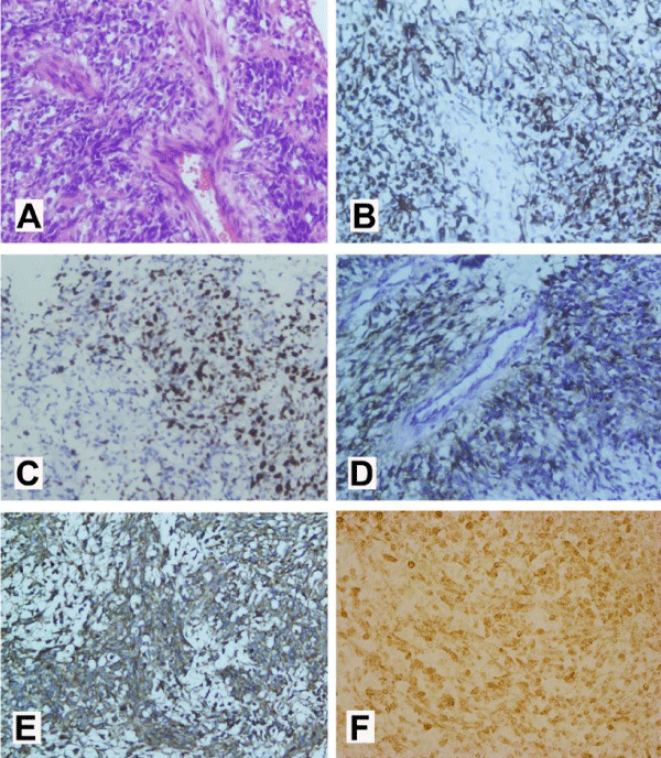
Figure 3:
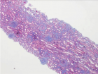
(A)Mitotic activity and perivascular pseudorosettes were conspicuous (hematoxylin and eosin, original magnification×200). (B) The tumor cells showed diffuse cytoplasmic glial fibrillary acidic protein (GFAP) immunoexpression (GFAP, original magnification×200), (C) diffuse nuclear Ki-67 immunoexpression (original magnification×200) and (D) diffuse perinuclear ‘‘dotlike’’ and ‘‘ring-like’’ epithelial membrane antigen (EMA) immunoexpression suggestive of ependymal differentiation (EMA, original magnification×200). (E) The tumor cells showed diffuse MMP-9 immunoexpression (MMP-9, original magnification×200); (F) The tumor cells showed diffuse L1CAM immunoexpression (L1CAM, original magnification×400).
Read Full Article HTML DOI: 10.29328/journal.acr.1001039 Cite this Article Read Full Article PDF
More Images
Similar Articles
-
Foley catheter balloon tamponade as a method of controlling iatrogenic pulmonary artery bleeding in redo thoracic surgeryMarcus Taylor*,Muhammad Asghar Nawaz,Ozhin Karadakhy,Denish Apparau,Kandadai Rammohan,Paul Waterworth. Foley catheter balloon tamponade as a method of controlling iatrogenic pulmonary artery bleeding in redo thoracic surgery. . 2019 doi: 10.29328/journal.acr.1001023; 3: 047-049
-
Acute and post burn reconstructive surgery of the female trunk with artificial dermis to facilitate healthy pregnancyDantzer E*,Campech G,Mazanovich M. Acute and post burn reconstructive surgery of the female trunk with artificial dermis to facilitate healthy pregnancy. . 2020 doi: 10.29328/journal.acr.1001036; 4: 028-031
-
Chronic subdural haematoma associated with arachnoid cyst of the middle fossa in a soccer player: Case report and review of the literatureElena Beretta*,Michele Incerti,Giuseppe Raudino,Gaspare F Montemagno,Franco Servadei. Chronic subdural haematoma associated with arachnoid cyst of the middle fossa in a soccer player: Case report and review of the literature. . 2020 doi: 10.29328/journal.acr.1001037; 4: 032-037
-
Ependymomas with extraneural metastasis to lung in children: A case report and literature reviewLiang Wang*,Miao Lou,Shunnan Ge,Guodong Gao,Yan Qu,Jiarui Zhang,Min Chao,Yang Jiao. Ependymomas with extraneural metastasis to lung in children: A case report and literature review. . 2020 doi: 10.29328/journal.acr.1001039; 4: 041-045
-
Exceptional intraoperative aspects of mesenteric venous gasWael Ferjaoui*,Mohamed Hajri,Aziz Atallah,Rached Bayar,Dhouha Bacha,Mohamed Tahar Khalfallah. Exceptional intraoperative aspects of mesenteric venous gas. . 2020 doi: 10.29328/journal.acr.1001042; 4: 050-051
-
Malignant transformation of an urachal cystWael Ferjaoui*,Dhouha Bacha,Rami Guizani,Aziz Atallah,Sana Ben Slama,Ahlem Lahmar. Malignant transformation of an urachal cyst. . 2020 doi: 10.29328/journal.acr.1001043; 4: 052-053
-
PET/MRI, aiming to improve the target for Fractionated Stereotactic Radiotherapy (FSRT) in recurrence of resected skull base meningioma after 2 years: Case reportSebastião Corrêa*. PET/MRI, aiming to improve the target for Fractionated Stereotactic Radiotherapy (FSRT) in recurrence of resected skull base meningioma after 2 years: Case report. . 2021 doi: 10.29328/journal.acr.1001044; 5: 001-003
-
Limb ischemia after coil migration used for a hypogastric aneurysm embolizationMiquel Gil Olaria*,Natalia Hernandez Wiesendange,Clàudia Riera Hernández,Carlos Esteban Gracia,Secundino Llagostera Pujol. Limb ischemia after coil migration used for a hypogastric aneurysm embolization. . 2021 doi: 10.29328/journal.acr.1001045; 5: 004-006
-
Influence of corneal spherical aberration, anterior chamber depth, and ocular axial length on the visual outcome with an extended depth of focus wavefront-designed intraocular lensBedei Andrea*,Carbonara Claudio,Farcomeni Alessio,Castellini Laura,Pietrelli Alessia. Influence of corneal spherical aberration, anterior chamber depth, and ocular axial length on the visual outcome with an extended depth of focus wavefront-designed intraocular lens. . 2022 doi: 10.29328/journal.acr.1001061; 6: 017-021
-
Neglected percutaneous rod extrusion following posterior occipitocervical instrumentation: a case reportMaximilian Reinhold*,Johannes Bonacker,Tobias Driesen,Wolfgang Lehmann. Neglected percutaneous rod extrusion following posterior occipitocervical instrumentation: a case report. . 2022 doi: 10.29328/journal.acr.1001063; 6: 024-026.
Recently Viewed
-
Minimising Carbon Footprint in Anaesthesia PracticeNisha Gandhi and Abinav Sarvesh SPS*. Minimising Carbon Footprint in Anaesthesia Practice. Int J Clin Anesth Res. 2024: doi: 10.29328/journal.ijcar.1001025; 8: 005-007
-
On Friedman equation, quadratic laws and the geometry of our universeS Kalimuthu*. On Friedman equation, quadratic laws and the geometry of our universe. Int J Phys Res Appl. 2021: doi: 10.29328/journal.ijpra.1001041; 4: 048-050
-
Texture of Thin Films of Aluminum Nitride Produced by Magnetron SputteringStrunin Vladimir Ivanovich,Baranova Larisa Vasilievna*,Baisova Bibigul Tulegenovna. Texture of Thin Films of Aluminum Nitride Produced by Magnetron Sputtering. Int J Phys Res Appl. 2025: doi: 10.29328/journal.ijpra.1001106; 8: 013-016
-
KYAMOS Software - Mini Review on the Computer-Aided Engineering IndustryAntonis Papadakis*,Sofia Nikolaidou. KYAMOS Software - Mini Review on the Computer-Aided Engineering Industry. Int J Phys Res Appl. 2025: doi: 10.29328/journal.ijpra.1001105; 8: 010-012
-
The Theory of ElementsAbdellah El-Mourabet*. The Theory of Elements. Int J Phys Res Appl. 2025: doi: 10.29328/journal.ijpra.1001104; 8: 001-009
Most Viewed
-
Evaluation of Biostimulants Based on Recovered Protein Hydrolysates from Animal By-products as Plant Growth EnhancersH Pérez-Aguilar*, M Lacruz-Asaro, F Arán-Ais. Evaluation of Biostimulants Based on Recovered Protein Hydrolysates from Animal By-products as Plant Growth Enhancers. J Plant Sci Phytopathol. 2023 doi: 10.29328/journal.jpsp.1001104; 7: 042-047
-
Sinonasal Myxoma Extending into the Orbit in a 4-Year Old: A Case PresentationJulian A Purrinos*, Ramzi Younis. Sinonasal Myxoma Extending into the Orbit in a 4-Year Old: A Case Presentation. Arch Case Rep. 2024 doi: 10.29328/journal.acr.1001099; 8: 075-077
-
Feasibility study of magnetic sensing for detecting single-neuron action potentialsDenis Tonini,Kai Wu,Renata Saha,Jian-Ping Wang*. Feasibility study of magnetic sensing for detecting single-neuron action potentials. Ann Biomed Sci Eng. 2022 doi: 10.29328/journal.abse.1001018; 6: 019-029
-
Pediatric Dysgerminoma: Unveiling a Rare Ovarian TumorFaten Limaiem*, Khalil Saffar, Ahmed Halouani. Pediatric Dysgerminoma: Unveiling a Rare Ovarian Tumor. Arch Case Rep. 2024 doi: 10.29328/journal.acr.1001087; 8: 010-013
-
Physical activity can change the physiological and psychological circumstances during COVID-19 pandemic: A narrative reviewKhashayar Maroufi*. Physical activity can change the physiological and psychological circumstances during COVID-19 pandemic: A narrative review. J Sports Med Ther. 2021 doi: 10.29328/journal.jsmt.1001051; 6: 001-007

HSPI: We're glad you're here. Please click "create a new Query" if you are a new visitor to our website and need further information from us.
If you are already a member of our network and need to keep track of any developments regarding a question you have already submitted, click "take me to my Query."


