Figure 1
Osteoclastic giant cell variant of urothelial carcinoma in a COVID- positive patient: A rare variant in an unusual circumstances
Chanchal Rana*, Divya Goel, Akanksha Singh, Suresh Babu and Vishwajeet Singh
Published: 13 April, 2021 | Volume 5 - Issue 1 | Pages: 009-011
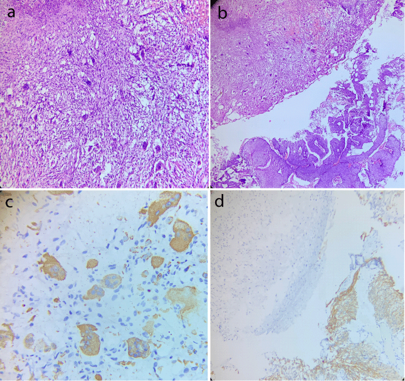
Figure 1:
Case of osteoblastic variant of bladder carcinoma displaying (a) Area rich in osteoblast like giant cells with intervening round to spindle shaped mononuclear cells (Hematoxylin and eosin stain, 400x); (b) Adjacent area of conventional high grade papillary urothelial carcinoma (Hematoxylin and eosin stain, 200x); (c) CD68 positivity in giant cell component and; (d) CK20 membranous positivity in conventional urothelial carcinoma component with absence of giant cell component.
Read Full Article HTML DOI: 10.29328/journal.acr.1001047 Cite this Article Read Full Article PDF
More Images
Similar Articles
-
Epstein-Barr infection causing toxic epidermal necrolysis, hemophagocytic lymphohistiocytosis and cerebritis in a pediatric patientAikaterini Solomou*,Vasileios Patriarcheas,Pantelis Kraniotis,Andreas Eliades. Epstein-Barr infection causing toxic epidermal necrolysis, hemophagocytic lymphohistiocytosis and cerebritis in a pediatric patient. . 2020 doi: 10.29328/journal.acr.1001032; 4: 015-019
-
Osteoclastic giant cell variant of urothelial carcinoma in a COVID- positive patient: A rare variant in an unusual circumstancesChanchal Rana*,Divya Goel,Akanksha Singh,Suresh Babu,Vishwajeet Singh. Osteoclastic giant cell variant of urothelial carcinoma in a COVID- positive patient: A rare variant in an unusual circumstances. . 2021 doi: 10.29328/journal.acr.1001047; 5: 009-011
-
Surgical management of splenic tuberculosis with pleural fistulation in a COVID-19 patientMohamed Firas Ayadi,Mohamed Hajri*,Ghofran Talbi,Hafedh Mestiri,Rached Bayar. Surgical management of splenic tuberculosis with pleural fistulation in a COVID-19 patient. . 2021 doi: 10.29328/journal.acr.1001052; 5: 027-027
-
Fatal acute necrotizing pancreatitis in a 15 years old boy, is it multisystem inflammatory syndrome in children associated with COVID-19; MIS-C?Masoumeh Asgarshirazi*,Khadije Daneshjou,Seyed Reza Raeeskarami,Mohammad Reza Keramati,Samrand Fattah Ghazi. Fatal acute necrotizing pancreatitis in a 15 years old boy, is it multisystem inflammatory syndrome in children associated with COVID-19; MIS-C?. . 2022 doi: 10.29328/journal.acr.1001056; 6: 001-004
-
Complex cyanotic congenital heart disease presenting as congenital heart block in a Nigerian infant: case report and literature reviewUjuanbi Amenawon Susan*,Amain Ebidimie Divine,Gregory Frances. Complex cyanotic congenital heart disease presenting as congenital heart block in a Nigerian infant: case report and literature review. . 2022 doi: 10.29328/journal.acr.1001058; 6: 009-012
-
A Case of X-Linked Hypophosphatemia: Exploring the Burden in a Single Family and the Significance of a Multidisciplinary ApproachAmrit Kaur Kaler, Nandini Shyamali Bora*, Kavyashree P, Ankita Nikam, Samrudhi Rane, Yash Tiwarekar, Shweta Limaye, Archana Juneja. A Case of X-Linked Hypophosphatemia: Exploring the Burden in a Single Family and the Significance of a Multidisciplinary Approach. . 2023 doi: 10.29328/journal.acr.1001076; 7: 042-045.
-
A Mini Review of Newly Identified Omicron SublineagesDasaradharami Reddy K*,Anusha S,Palem Chandrakala. A Mini Review of Newly Identified Omicron Sublineages. . 2023 doi: 10.29328/journal.acr.1001082; 7: 066-076
-
Towards A 21st Century Systematize the Ideas; COVID-19, Sustainability and Discourse of SDG, (Sustainable Development Goals), The Cities and Housing ModelsHülya Coskun*. Towards A 21st Century Systematize the Ideas; COVID-19, Sustainability and Discourse of SDG, (Sustainable Development Goals), The Cities and Housing Models. . 2024 doi: 10.29328/journal.acr.1001089; 8: 027-035
-
Post-COVID-19 Era, 15th Minutes City New Urban Model Changing Housing Design and ModelsHülya Coskun*. Post-COVID-19 Era, 15th Minutes City New Urban Model Changing Housing Design and Models. . 2024 doi: 10.29328/journal.acr.1001098; 8: 063-074
-
Is Acupuncture Efficient for Treating Long COVID? Case ReportsDavid Lake*, Philippe Poindron. Is Acupuncture Efficient for Treating Long COVID? Case Reports. . 2024 doi: 10.29328/journal.acr.1001107; 8: 110-115
Recently Viewed
-
Autoantibodies in Autoimmune Addison’s Disease: Why are they Important?Maria Rosaria De Cagna, Norma Notaristefano, Maurizio Schiavone, Gianluca Palatella, Federica Ranù, Carmela Presicci, Valerio Cecinati, Marilina Tampoia*. Autoantibodies in Autoimmune Addison’s Disease: Why are they Important?. Arch Pathol Clin Res. 2024: doi: 10.29328/journal.apcr.1001042; 8: 012-015
-
Hepatic Pseudolymphoma Mimicking Neoplasia in Primary Biliary Cholangitis: A Case ReportJeremy Hassoun,Aurélie Bornand,Alexis Ricoeur,Giulia Magini,Nicolas Goossens,Laurent Spahr*. Hepatic Pseudolymphoma Mimicking Neoplasia in Primary Biliary Cholangitis: A Case Report. Arch Case Rep. 2024: doi: 10.29328/journal.acr.1001115; 8: 152-155
-
Statistical and equation model analysis on COVID-19Bin Zhao*,Jinming Cao. Statistical and equation model analysis on COVID-19. Arch Biotechnol Biomed. 2020: doi: 10.29328/journal.abb.1001016; 4: 005-012
-
Sinonasal Myxoma Extending into the Orbit in a 4-Year Old: A Case PresentationJulian A Purrinos*, Ramzi Younis. Sinonasal Myxoma Extending into the Orbit in a 4-Year Old: A Case Presentation. Arch Case Rep. 2024: doi: 10.29328/journal.acr.1001099; 8: 075-077
-
Investigation of Stain Patterns from Diverse Blood Samples on Various SurfacesSonia Rajkumari*. Investigation of Stain Patterns from Diverse Blood Samples on Various Surfaces. J Forensic Sci Res. 2024: doi: 10.29328/journal.jfsr.1001061; 8: 028034
Most Viewed
-
Evaluation of Biostimulants Based on Recovered Protein Hydrolysates from Animal By-products as Plant Growth EnhancersH Pérez-Aguilar*, M Lacruz-Asaro, F Arán-Ais. Evaluation of Biostimulants Based on Recovered Protein Hydrolysates from Animal By-products as Plant Growth Enhancers. J Plant Sci Phytopathol. 2023 doi: 10.29328/journal.jpsp.1001104; 7: 042-047
-
Sinonasal Myxoma Extending into the Orbit in a 4-Year Old: A Case PresentationJulian A Purrinos*, Ramzi Younis. Sinonasal Myxoma Extending into the Orbit in a 4-Year Old: A Case Presentation. Arch Case Rep. 2024 doi: 10.29328/journal.acr.1001099; 8: 075-077
-
Feasibility study of magnetic sensing for detecting single-neuron action potentialsDenis Tonini,Kai Wu,Renata Saha,Jian-Ping Wang*. Feasibility study of magnetic sensing for detecting single-neuron action potentials. Ann Biomed Sci Eng. 2022 doi: 10.29328/journal.abse.1001018; 6: 019-029
-
Pediatric Dysgerminoma: Unveiling a Rare Ovarian TumorFaten Limaiem*, Khalil Saffar, Ahmed Halouani. Pediatric Dysgerminoma: Unveiling a Rare Ovarian Tumor. Arch Case Rep. 2024 doi: 10.29328/journal.acr.1001087; 8: 010-013
-
Physical activity can change the physiological and psychological circumstances during COVID-19 pandemic: A narrative reviewKhashayar Maroufi*. Physical activity can change the physiological and psychological circumstances during COVID-19 pandemic: A narrative review. J Sports Med Ther. 2021 doi: 10.29328/journal.jsmt.1001051; 6: 001-007

HSPI: We're glad you're here. Please click "create a new Query" if you are a new visitor to our website and need further information from us.
If you are already a member of our network and need to keep track of any developments regarding a question you have already submitted, click "take me to my Query."


