Figure 2
Oncocytic papillary cystadenoma of right laryngeal ventricle
Tomasz Ścięgosz*, Renata Kwiatek, Izabela El-Hassanieh and Piotr Ziółkowski
Published: 30 April, 2021 | Volume 5 - Issue 1 | Pages: 012-013
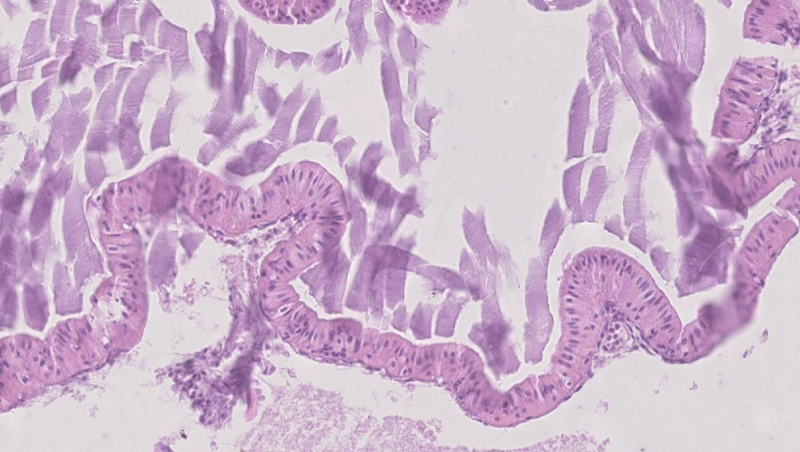
Figure 2:
The cysts were lined with two layers of oncocytic cells; the continuous luminal layer of columnar cells with palisading cigar-shaped nuclei and the intermittent layer of basal cells with round nuclei. HE staining, 200x.
Read Full Article HTML DOI: 10.29328/journal.acr.1001048 Cite this Article Read Full Article PDF
More Images
Similar Articles
-
Clinical, histopathological and surgical evaluations of persistent oropharyngeal membrane case in a calfVehbi Gunes*,Gultekin Atalan,Latife Cakir Bayram,Kemal Varol,Hanifi Erol,Ihsan Keles,Ali C Onmaz. Clinical, histopathological and surgical evaluations of persistent oropharyngeal membrane case in a calf. . 2019 doi: 10.29328/journal.acr.1001016; 3: 021-025
-
Epiphora as a sign of unexpected underlying squamous cell carcinoma within sinonasal inverted papillomaFilippo Confalonieri*,Alessandra Di Maria,Raffaele Piscopo,Laura Balia,Luca Malvezzi. Epiphora as a sign of unexpected underlying squamous cell carcinoma within sinonasal inverted papilloma. . 2020 doi: 10.29328/journal.acr.1001038; 4: 038-040
-
Oncocytic papillary cystadenoma of right laryngeal ventricleTomasz Ścięgosz*,Renata Kwiatek,Izabela El-Hassanieh,Piotr Ziółkowski. Oncocytic papillary cystadenoma of right laryngeal ventricle. . 2021 doi: 10.29328/journal.acr.1001048; 5: 012-013
-
Endoscopic Endonasal total Removal of a Suprasellar, Preinfundibular Retro Chiasmatic Craniopharyngioma: A Surgical Case ReportAlessandra Alfieri, Armando Rapanà, Ferdinando Caranci. Endoscopic Endonasal total Removal of a Suprasellar, Preinfundibular Retro Chiasmatic Craniopharyngioma: A Surgical Case Report. . 2024 doi: 10.29328/journal.acr.1001090; 8: 036-038
-
Sinonasal Myxoma Extending into the Orbit in a 4-Year Old: A Case PresentationJulian A Purrinos*, Ramzi Younis. Sinonasal Myxoma Extending into the Orbit in a 4-Year Old: A Case Presentation. . 2024 doi: 10.29328/journal.acr.1001099; 8: 075-077
Recently Viewed
-
String Theory without Extra Dimensions and Without SupersymmetryBassem Aouadi*. String Theory without Extra Dimensions and Without Supersymmetry. Int J Phys Res Appl. 2024: doi: 10.29328/journal.ijpra.1001103; 7: 162-166
-
Fabrication and Optimization of Alginate Membranes for Improved Wastewater TreatmentJohn Sunday Uzochukwu,Nweke Chinenyenwa Nkeiruka*,Nwachukwu Josiah Odinaka,Olufemi Gideon Olajide. Fabrication and Optimization of Alginate Membranes for Improved Wastewater Treatment. Arch Case Rep. 2025: doi: ; 9: 026-043
-
Navigating Weight Management with Stevia: Insights into Glycemic ControlShashikanth Kharat*, Sanjana Mali*. Navigating Weight Management with Stevia: Insights into Glycemic Control. New Insights Obes Gene Beyond. 2024: doi: 10.29328/journal.niogb.1001021; 8: 006-008
-
Convalescent plasma therapy in aHUS patient with SARS-CoV-2 infectionEmma Diletta Stea*,Virginia Pronzo,Francesco Pesce,Marco Fiorentino,Adele Mitrotti,Vincenzo Di Leo,Cosma Cortese,Annalisa Casanova,Sebastiano Nestola,Flavia Capaccio,Loreto Gesualdo. Convalescent plasma therapy in aHUS patient with SARS-CoV-2 infection. J Clini Nephrol. 2022: doi: 10.29328/journal.jcn.1001088; 6: 036-039
-
An Uncommon Case Report of Hypothyroidism, Type 1 Diabetes Mellitus, and Systemic Lupus Erythematosus with an Immunosuppressive Consequence: A Case ReportArif Hoda*, Shruti R Shinde, Avinash Chaudhari, Sameer Vyahalkar, Amar Kulkarni, Pooja Binani, Amit Nagrik. An Uncommon Case Report of Hypothyroidism, Type 1 Diabetes Mellitus, and Systemic Lupus Erythematosus with an Immunosuppressive Consequence: A Case Report. J Clini Nephrol. 2024: doi: 10.29328/journal.jcn.1001138; 8: 118-123
Most Viewed
-
Evaluation of Biostimulants Based on Recovered Protein Hydrolysates from Animal By-products as Plant Growth EnhancersH Pérez-Aguilar*, M Lacruz-Asaro, F Arán-Ais. Evaluation of Biostimulants Based on Recovered Protein Hydrolysates from Animal By-products as Plant Growth Enhancers. J Plant Sci Phytopathol. 2023 doi: 10.29328/journal.jpsp.1001104; 7: 042-047
-
Sinonasal Myxoma Extending into the Orbit in a 4-Year Old: A Case PresentationJulian A Purrinos*, Ramzi Younis. Sinonasal Myxoma Extending into the Orbit in a 4-Year Old: A Case Presentation. Arch Case Rep. 2024 doi: 10.29328/journal.acr.1001099; 8: 075-077
-
Feasibility study of magnetic sensing for detecting single-neuron action potentialsDenis Tonini,Kai Wu,Renata Saha,Jian-Ping Wang*. Feasibility study of magnetic sensing for detecting single-neuron action potentials. Ann Biomed Sci Eng. 2022 doi: 10.29328/journal.abse.1001018; 6: 019-029
-
Pediatric Dysgerminoma: Unveiling a Rare Ovarian TumorFaten Limaiem*, Khalil Saffar, Ahmed Halouani. Pediatric Dysgerminoma: Unveiling a Rare Ovarian Tumor. Arch Case Rep. 2024 doi: 10.29328/journal.acr.1001087; 8: 010-013
-
Physical activity can change the physiological and psychological circumstances during COVID-19 pandemic: A narrative reviewKhashayar Maroufi*. Physical activity can change the physiological and psychological circumstances during COVID-19 pandemic: A narrative review. J Sports Med Ther. 2021 doi: 10.29328/journal.jsmt.1001051; 6: 001-007

HSPI: We're glad you're here. Please click "create a new Query" if you are a new visitor to our website and need further information from us.
If you are already a member of our network and need to keep track of any developments regarding a question you have already submitted, click "take me to my Query."


