Figure 1
Uncommon first diagnosis of metastatic papillary thyroid carcinoma with “signet-ring” cells morphology through pericardial effusion
Angeliki Cheva, Sokratis Tsagkaropoulos*, Panagiotis Pepis, Antonia Syrnioti and Christoforos Foroulis
Published: 20 January, 2022 | Volume 6 - Issue 1 | Pages: 005-008
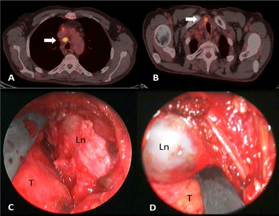
Figure 1:
PET/CT scan revealing high metabolic activity in right paratracheal (A) and pretracheal (B) lymph nodes. Introperative findings corresponding to the PET/CT scan images (C, D). LN: lymph nodes, T: trachea.
Read Full Article HTML DOI: 10.29328/journal.acr.1001057 Cite this Article Read Full Article PDF
More Images
Similar Articles
-
Uncommon first diagnosis of metastatic papillary thyroid carcinoma with “signet-ring” cells morphology through pericardial effusionAngeliki Cheva,Sokratis Tsagkaropoulos*,Panagiotis Pepis,Antonia Syrnioti,Christoforos Foroulis. Uncommon first diagnosis of metastatic papillary thyroid carcinoma with “signet-ring” cells morphology through pericardial effusion. . 2022 doi: 10.29328/journal.acr.1001057; 6: 005-008
Recently Viewed
-
Various Theories of Fast and Ultrafast Magnetization DynamicsManfred Fähnle*. Various Theories of Fast and Ultrafast Magnetization Dynamics. Int J Phys Res Appl. 2024: doi: 10.29328/journal.ijpra.1001101; 7: 154-158
-
Forest History Association of WisconsinEd Bauer*. Forest History Association of Wisconsin. J Radiol Oncol. 2024: doi: 10.29328/journal.jro.1001071; 8: 093-096
-
Synthesis of Carbon Nano Fiber from Organic Waste and Activation of its Surface AreaHimanshu Narayan*,Brijesh Gaud,Amrita Singh,Sandesh Jaybhaye. Synthesis of Carbon Nano Fiber from Organic Waste and Activation of its Surface Area. Int J Phys Res Appl. 2019: doi: 10.29328/journal.ijpra.1001017; 2: 056-059
-
Obesity Surgery in SpainAniceto Baltasar*. Obesity Surgery in Spain. New Insights Obes Gene Beyond. 2020: doi: 10.29328/journal.niogb.1001013; 4: 013-021
-
Tamsulosin and Dementia in old age: Is there any relationship?Irami Araújo-Filho*,Rebecca Renata Lapenda do Monte,Karina de Andrade Vidal Costa,Amália Cinthia Meneses Rêgo. Tamsulosin and Dementia in old age: Is there any relationship?. J Neurosci Neurol Disord. 2019: doi: 10.29328/journal.jnnd.1001025; 3: 145-147
Most Viewed
-
Evaluation of Biostimulants Based on Recovered Protein Hydrolysates from Animal By-products as Plant Growth EnhancersH Pérez-Aguilar*, M Lacruz-Asaro, F Arán-Ais. Evaluation of Biostimulants Based on Recovered Protein Hydrolysates from Animal By-products as Plant Growth Enhancers. J Plant Sci Phytopathol. 2023 doi: 10.29328/journal.jpsp.1001104; 7: 042-047
-
Sinonasal Myxoma Extending into the Orbit in a 4-Year Old: A Case PresentationJulian A Purrinos*, Ramzi Younis. Sinonasal Myxoma Extending into the Orbit in a 4-Year Old: A Case Presentation. Arch Case Rep. 2024 doi: 10.29328/journal.acr.1001099; 8: 075-077
-
Feasibility study of magnetic sensing for detecting single-neuron action potentialsDenis Tonini,Kai Wu,Renata Saha,Jian-Ping Wang*. Feasibility study of magnetic sensing for detecting single-neuron action potentials. Ann Biomed Sci Eng. 2022 doi: 10.29328/journal.abse.1001018; 6: 019-029
-
Pediatric Dysgerminoma: Unveiling a Rare Ovarian TumorFaten Limaiem*, Khalil Saffar, Ahmed Halouani. Pediatric Dysgerminoma: Unveiling a Rare Ovarian Tumor. Arch Case Rep. 2024 doi: 10.29328/journal.acr.1001087; 8: 010-013
-
Physical activity can change the physiological and psychological circumstances during COVID-19 pandemic: A narrative reviewKhashayar Maroufi*. Physical activity can change the physiological and psychological circumstances during COVID-19 pandemic: A narrative review. J Sports Med Ther. 2021 doi: 10.29328/journal.jsmt.1001051; 6: 001-007

HSPI: We're glad you're here. Please click "create a new Query" if you are a new visitor to our website and need further information from us.
If you are already a member of our network and need to keep track of any developments regarding a question you have already submitted, click "take me to my Query."


