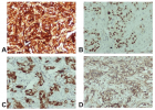
Figure 2
Uncommon first diagnosis of metastatic papillary thyroid carcinoma with “signet-ring” cells morphology through pericardial effusion
Angeliki Cheva, Sokratis Tsagkaropoulos*, Panagiotis Pepis, Antonia Syrnioti and Christoforos Foroulis
Published: 20 January, 2022 | Volume 6 - Issue 1 | Pages: 005-008
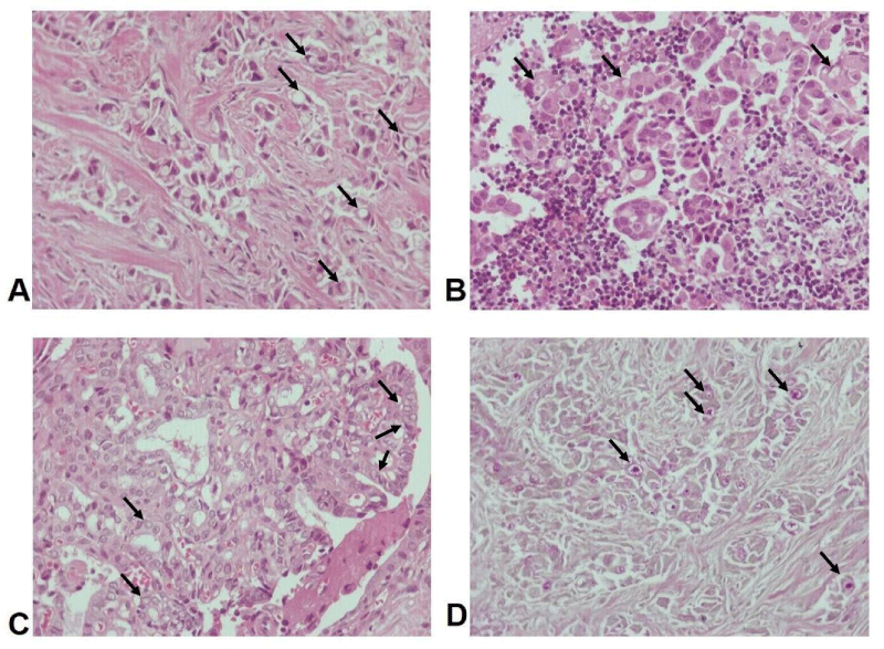
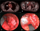
Figure 2:
A, B. Hematoxylin and eosin x200: “Signet-ring” cell morphology of neoplastic cells in pericardial tissue (A) and lymph node (B), C. “Empty” nuclei with grooving membrane and small inconspicuous nucleoli with papillary and follicular growth pattern in pericardial tissue, D. Periodic Acid-Schiff (PAS) x200, revealing the presence of intracytoplasmic music. Arrows indicate pathologic cells.
Read Full Article HTML DOI: 10.29328/journal.acr.1001057 Cite this Article Read Full Article PDF
More Images
Similar Articles
-
Uncommon first diagnosis of metastatic papillary thyroid carcinoma with “signet-ring” cells morphology through pericardial effusionAngeliki Cheva,Sokratis Tsagkaropoulos*,Panagiotis Pepis,Antonia Syrnioti,Christoforos Foroulis. Uncommon first diagnosis of metastatic papillary thyroid carcinoma with “signet-ring” cells morphology through pericardial effusion. . 2022 doi: 10.29328/journal.acr.1001057; 6: 005-008
Recently Viewed
-
Role of novel cardiac biomarkers for the diagnosis, risk stratification, and prognostication among patients with heart failureJennifer Miao,Joel Estis,Yan Ru Su,John A Todd,Daniel J Lenihan*. Role of novel cardiac biomarkers for the diagnosis, risk stratification, and prognostication among patients with heart failure. J Cardiol Cardiovasc Med. 2019: doi: 10.29328/journal.jccm.1001049; 4: 103-109
-
Late discover of a traumatic cardiac injury: Case reportBenlafqih C,Bouhdadi H*,Bakkali A,Rhissassi J,Sayah R,Laaroussi M. Late discover of a traumatic cardiac injury: Case report. J Cardiol Cardiovasc Med. 2019: doi: 10.29328/journal.jccm.1001048; 4: 100-102
-
Timing of cardiac surgery and other intervention among children with congenital heart disease: A review articleChinawa JM*,Adiele KD,Ujunwa FA,Onukwuli VO,Arodiwe I,Chinawa AT,Obidike EO,Chukwu BF. Timing of cardiac surgery and other intervention among children with congenital heart disease: A review article. J Cardiol Cardiovasc Med. 2019: doi: 10.29328/journal.jccm.1001047; 4: 094-099
-
P wave dispersion in patients with premenstrual dysphoric disorderSeyda Yavuzkir,Suna Aydin*,Melike Baspinar,Sevda Korkmaz,Murad Atmaca,Rulin Deniz,Yakup Baykus,Mustafa Yavuzkir. P wave dispersion in patients with premenstrual dysphoric disorder. J Cardiol Cardiovasc Med. 2019: doi: 10.29328/journal.jccm.1001046; 4: 090-093
-
Preclinical stiff heart is a marker of cardiovascular morbimortality in apparently healthy populationCharles Fauvel,Michael Bubenheim,Olivier Raitière,Charlotte Vallet,Nassima Si Belkacem,Fabrice Bauer*. Preclinical stiff heart is a marker of cardiovascular morbimortality in apparently healthy population. J Cardiol Cardiovasc Med. 2019: doi: 10.29328/journal.jccm.1001045; 4: 083-089
Most Viewed
-
Evaluation of Biostimulants Based on Recovered Protein Hydrolysates from Animal By-products as Plant Growth EnhancersH Pérez-Aguilar*, M Lacruz-Asaro, F Arán-Ais. Evaluation of Biostimulants Based on Recovered Protein Hydrolysates from Animal By-products as Plant Growth Enhancers. J Plant Sci Phytopathol. 2023 doi: 10.29328/journal.jpsp.1001104; 7: 042-047
-
Sinonasal Myxoma Extending into the Orbit in a 4-Year Old: A Case PresentationJulian A Purrinos*, Ramzi Younis. Sinonasal Myxoma Extending into the Orbit in a 4-Year Old: A Case Presentation. Arch Case Rep. 2024 doi: 10.29328/journal.acr.1001099; 8: 075-077
-
Feasibility study of magnetic sensing for detecting single-neuron action potentialsDenis Tonini,Kai Wu,Renata Saha,Jian-Ping Wang*. Feasibility study of magnetic sensing for detecting single-neuron action potentials. Ann Biomed Sci Eng. 2022 doi: 10.29328/journal.abse.1001018; 6: 019-029
-
Pediatric Dysgerminoma: Unveiling a Rare Ovarian TumorFaten Limaiem*, Khalil Saffar, Ahmed Halouani. Pediatric Dysgerminoma: Unveiling a Rare Ovarian Tumor. Arch Case Rep. 2024 doi: 10.29328/journal.acr.1001087; 8: 010-013
-
Physical activity can change the physiological and psychological circumstances during COVID-19 pandemic: A narrative reviewKhashayar Maroufi*. Physical activity can change the physiological and psychological circumstances during COVID-19 pandemic: A narrative review. J Sports Med Ther. 2021 doi: 10.29328/journal.jsmt.1001051; 6: 001-007

HSPI: We're glad you're here. Please click "create a new Query" if you are a new visitor to our website and need further information from us.
If you are already a member of our network and need to keep track of any developments regarding a question you have already submitted, click "take me to my Query."


